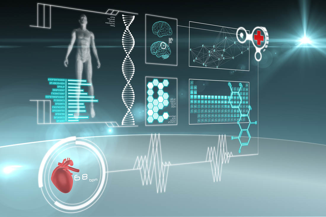
In the past century, astounding contributions to the biomedical imaging field have continuously improved the ability to look inside the human body non-invasively.
X-rays were the only means to do it initially. But other energy forms, which include ultrasound and electromagnetic fields, have evolved imaging from being used only for patient care to the studies of biological structure and function.
 |
| Image source: engineering.case.edu |
Low-quality images are also a thing of the past. New technology has provided high-definition and three-dimensional images, and in some instances, videos. These have shaped how diagnosis and treatment are being performed.
Considering the proximity of imaging technology to the practical limits of spatial resolution in all modalities, improving image quality is no longer the major driver of innovation in imaging.
What physicists are striving to achieve now is a reduction in radiation dosage to minimize the patient’s exposure to its harmful effects. The medical field had already come a long way from the early limitations of X-ray when radiation exposure was much higher than it was today. Still, it is a legitimate concern.
 |
| Image source: ultrasoundschoolsinfo.com |
Efficiency of resource utilization is also an objective of technical innovation. A significant reduction in cost is seen to make it more accessible to more people.
Additionally, the scarcest of resources – time – needs to be saved by speeding up the scan or imaging time to provide results as early as possible. This will benefit not only patients but also the physicians and medical workers.
Akash Monpara is currently a student in John Hopkins University, majoring in biomedical engineering. Connect with this Google+ account to read more about the industry.